525 Amherst St., Suite 102 • Winchester, VA 22601 • P 540-667-5535 • F 540-667-5536

Cataract Procedures
Understanding Your Cataract Procedures
Your eye has a clear lens, just like a camera. Age, injury, or other factors can cause this lens to become clouded, into what is known as a cataract. Normally, a clear lens provides clear vision. A cataract lens blurs, dims, discolors, and clouds one’s vision. Surgery removes this cataract, and replaces it with a small, clear plastic lens. With a clear normal lens, the images are focused clearly on the retina. Vision is clear. With a cataract, the lens is cloudy, causing the image to become blurred and yellowed. Vision is hazy and colors become faded.Types of Cataracts
There are many types of cataracts. As cataracts can develop slowly over a long period of time, significant decreased vision may not be noticed. In some, only part of the lens is affected, and yet, this can cause a great loss in the quality of vision. The replacement intraocular lenses are technically refined and versatile.Treatment Overview
For most cases, a small incision is made in the front part of the eye, and the cataract lens is removed using a special ultrasonic probe. The bag holding the lens is left intact. The new lens is emplaced in this bag. Sometimes, following cataract surgery, the lens bag can develop opacities behind the implanted lens, called a secondary cataract. Laser treatment at a later date creates a hole in the bag to restore clear vision.Cataract Surgery
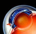
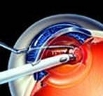
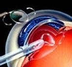
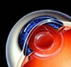
The surgery begins with a very small incision, in the white sclera of the eye, or near the edge of the clear cornea. A special protective viscoelastic material, or air, is placed inside the eye. The lens bag is opened on top. Fluid is used to separate the lens from its outer bag - this is called hydrodissection. Next, a special ultrasonic probe is used, to break up and remove the lens - this is called phacoemulsification. The natural lens bag is left intact. It is polished and cleaned, using irrigation and aspiration. In most cases, the folded intraocular lens is placed through the small incision, where it is unfolded into the lens bag. 1. A very small "No Stitch" incision is made in the side of the cornea. Such an incision promotes fast and more comfortable recovery. 2. The bag of the lens is opened and a special ultrasonic probe (phacoemulsifier) removes the cloudy lens. 3. A small foldable artificial lens is inserted through the small incision to replace the cataract lens. 4. The final replacement lens is shown in place. It is not required to suture the small "No Stitch" incision. This modern approach usually requires no suture for the very small incision, and the surgery is complete. The computerized cataract ultrasonic system provides total surgeon control of both intraocular pressure and cataract extraction. Other techniques may be used in special cases.The Result
Before cataract surgery, the lens is cloudy, causing images to be blurred and yellowed. Vision is hazy and colors are faded. After surgery, the new replacement lens provides a clear and focused image on the retina. Vision is clear.Summary
Cataracts mostly affect people over 60 years of age, but they can occur at any age. Most cataract surgeries are an out-patient procedure, taking just a few minutes, returning to most daily activities within a couple of days. Cataract surgery, as with all surgeries, does contain an element of risk which must be fully considered and discussed with your doctor. Click here to go back to our services page

The Winchester Eye
Surgery Center is staffed
by caring professionals to
serve patients' surgical
needs and provide
cataract, corneal,
oculoplastic, glaucoma
and general
ophthalmology
procedures.